what protein complex produces the proton gradient used to power the product of atp
Introduction
The electron transport chain is a series of four protein complexes that couple redox reactions, creating an electrochemical gradient that leads to the creation of ATP in a consummate arrangement named oxidative phosphorylation. It occurs in mitochondria in both cellular respiration and photosynthesis. In the former, the electrons come from breaking downward organic molecules, and free energy is released. In the latter, the electrons enter the chain afterward being excited by low-cal, and the energy released is used to build carbohydrates.
Fundamentals
Aerobic cellular respiration is made up of three parts: glycolysis, the citric acrid (Krebs) cycle, and oxidative phosphorylation. In glycolysis, glucose metabolizes into 2 molecules of pyruvate, with an output of ATP and nicotinamide adenine dinucleotide (NADH). Each pyruvate oxidizes into acetyl CoA and an boosted molecule of NADH and carbon dioxide (CO2). The acetyl CoA is then used in the citric acid cycle, which is a concatenation of chemic reactions that produce CO2, NADH, flavin adenine dinucleotide (FADH2), and ATP. In the final stride, the iii NADH and ane FADH2 amassed from the previous steps are used in oxidative phosphorylation, to make water and ATP.
Oxidative phosphorylation has two parts: the electron transport chain (ETC) and chemiosmosis. The ETC is a collection of proteins spring to the inner mitochondrial membrane and organic molecules, which electrons pass through in a series of redox reactions, and release energy. The free energy released forms a proton gradient, which is used in chemiosmosis to make a large amount of ATP by the poly peptide ATP-synthase.
Photosynthesis is a metabolic process that converts light energy into chemical energy to build sugars. In the light-dependent reactions, light energy and h2o are used to make ATP, NADPH, and oxygen (O2). The proton gradient used to make the ATP forms via an electron ship chain. In the lite-independent reactions, sugar is made from the ATP and NADPH from the previous reactions.
Cellular
In the electron ship concatenation (ETC), the electrons go through a chain of proteins that increases its reduction potential and causes a release in energy. Most of this energy is dissipated as heat or utilized to pump hydrogen ions (H+) from the mitochondrial matrix to the intermembrane space and create a proton gradient. This slope increases the acidity in the intermembrane space and creates an electric difference with a positive charge outside and a negative accuse inside. The ETC proteins in a general order are complex I, circuitous II, coenzyme Q, complex III, cytochrome C, and complex Iv.
- Circuitous I, likewise known as ubiquinone oxidoreductase, is fabricated up of NADH dehydrogenase, flavin mononucleotide (FMN), and viii iron-sulfur (Fe-South) clusters. The NADH donated from glycolysis, and the citric acid cycle is oxidized hither, transferring ii electrons from NADH to FMN. And then they are transferred to the Iron-Southward clusters and finally from Atomic number 26-Southward to coenzyme Q. During this procedure, 4 hydrogen ions pass from the mitochondrial matrix to the intermembrane space, contributing to the electrochemical slope. Complex I may also play an important role in causing apoptosis in programmed cell expiry.[1][2][3][4][one]
- (NADH + H+) + CoQ + 4 H+(matrix) -> NAD+ + CoQH2 + 4 H+(intermembrane)
- Complex Two, also known as succinate dehydrogenase, accepts electrons from succinate (an intermediate in the citric acrid cycle) and acts as a second entry bespeak to the ETC. When succinate oxidizes to fumarate, 2 electrons are accepted by FAD within complex Two. FAD passes them to Fe-S clusters and and then to coenzyme Q, similar to circuitous I. However; no protons are translocated beyond the membrane by circuitous 2, therefore less ATP is produced with this pathway.[five][6]
- Succinate + FAD -> Fumarate + 2 H+(matrix) + FADH2
- FADH2 + CoQ -> FAD + CoQH2
- Glycerol-3-Phosphate dehydrogenase and Acyl-CoA dehydrogenase also have electrons from glycerol-iii-P and fat acyl-CoA, respectively. Inclusion of these poly peptide complexes allows for the donation to the ETC by cytosolic NADH (glycerol-3-P acts as a shuttle to regenerate cytosolic NAD from NADH) and fatty acids undergoing beta-oxidation within the mitochondria (acyl-CoA is oxidized to enoyl-CoA in the first footstep, producing FADH2).[7][viii]
- Coenzyme Q, likewise known equally ubiquinone (CoQ), is made up of quinone and a hydrophobic tail. Its purpose is to part as an electron carrier and transfer electrons to complex III. Coenzyme Q undergoes reduction to semiquinone (partially reduced, radical course CoQH-) and ubiquinol (fully reduced CoQH2) through the Q wheel. This process receives further elaboration under Complex Three.
- Circuitous 3, too known as cytochrome c reductase, is made up of cytochrome b, Rieske subunits (containing two Fe-S clusters), and cytochrome c proteins. A cytochrome is a protein involved in electron transfer that contains a heme group. The heme groups alternating betwixt ferrous (Fe2+) and ferric (Fe3+) states during the electron transfer. Because cytochrome c can only have a unmarried electron at a time, this process occurs in two steps (the Q wheel), in dissimilarity to the single-step complex I and II pathways. Circuitous III also releases 4 protons into the intermembrane infinite at the end of a full Q cycle, contributing to the slope. Cytochrome c then transfers the electrons 1 at a time to complex Iv.[9][ten][11]
- Q Bike:
- Step one in the Q cycle involves ubiquinol (CoQH2) and ubiquinone (CoQ) binding to ii separate sites on circuitous III. CoQH2 transfers each electron to a different path. One electron goes to Iron-Southward and so cytochrome c, while the 2d electron is transferred to cytochrome b and so to CoQ bound at the other site. While this occurs, 2 H+ ions are released into the intermembrane infinite, contributing to the proton gradient. CoQH2 is now oxidized to ubiquinone and dissociates from the complex. The CoQ bound at the second site enters a transitional CoQH- radical state from accepting ane of the electrons.
- The second pace of the cycle involves a repeat of the offset: a new CoQH2 binds to the kickoff site and transfers two electrons similar before (and 2 more H+ ions released). Again, i electron passes to cytochrome c and one to cytochrome b, which this time works to reduce CoQH- to CoQH2 before it dissociates from complex III and can be recycled. In this style, one full cycle appears as follows:[12]
- 2 CoQH2(site i) + CoQ(site 2) + 2 Cyt c(ox) + 2 H+(matrix) -> 2 CoQ(site 1) + CoQH2(site 2) + two Cyt c(cherry-red) + 4 H+(intermembrane)
- Q Bike:
- Circuitous Four, also known as cytochrome c oxidase, oxidizes cytochrome c and transfers the electrons to oxygen, the final electron carrier in aerobic cellular respiration. The cytochrome proteins a and a3, in improver to heme and copper groups in circuitous IV transfer the donated electrons to the bound dioxygen species, converting it into molecules of h2o. The gratuitous free energy from the electron transfer causes 4 protons to move into the intermembrane space contributing to the proton gradient. Oxygen reduces via the following reaction:[thirteen][14]
- ii cytochrome c(crimson) + ½O2 + 4 H+(matrix) -> 2 cytochrome c(ox) + one Water + two H+(intermembrane)
ATP synthase, also called complex 5, uses the ETC generated proton gradient across the inner mitochondrial membrane to class ATP. ATP-synthase contains up of F0 and F1 subunits, which act as a rotational motor system. F0 is hydrophobic and embedded in the inner mitochondrial membrane. It contains a proton corridor that is protonated and deprotonated repeatedly equally H+ ions flow down the gradient from intermembrane space to matrix. The alternate ionization of F0 causes rotation, which alters the orientation of the F1 subunits. F1 is hydrophilic and faces the mitochondrial matrix. Conformational changes in F1 subunits catalyze the formation of ATP from ADP and Pi. For every 4 H+ ions, 1 ATP is produced. ATP-synthase can also be forced to run in opposite, consuming ATP to produce a hydrogen gradient, as is seen in some bacteria.[15][16][17]
Molecular
Nicotinamide adenine dinucleotide has two forms: NAD+ (oxidized) and NADH (reduced). Information technology is a dinucleotide connected by phosphate groups. Ane nucleoside has an adenine base of operations and the other nicotinamide. When involved in metabolic redox reactions, the machinery is as shown in Reaction 1.
- Reaction i: RH2 + NAD+ -> R + H+ + NADH
R is the reactant, for example, sugar.
NADH enters the ETC at complex I and produces a total of 10 H+ ions through the ETC (four from circuitous I, 4 from complex 3, and 2 from complex Iv). ATP-synthase synthesizes ane ATP for four H+ ions. Therefore, i NADH = 10 H+, and 10/four H+ per ATP = two.5 ATP per NADH (**some sources round up**). When NADH is oxidized, information technology breaks into NAD+, H+, and 2 east- as shown in Reaction 2.
- Reaction 2: NADH -> H+ + NAD+ + 2 e-
Flavin adenine dinucleotide has four redox states, three of them being FAD (quinone, fully oxidized form), FADH- (semiquinone, partially oxidized), and FADH2 (hydroquinone, fully reduced). FAD is made up of an adenine nucleotide and a flavin mononucleotide (FMN), connected by phosphate groups. FMN is synthesized in role from vitamin B2 (riboflavin). FAD contains a highly stable aromatic band, and FADH2 does non. When FADH2 oxidizes, information technology becomes aromatic and releases energy, as seen in Reaction 3. This state makes FAD a potent oxidizing agent, with an even more than positive reduction potential than NAD. FADH2 enters the ETC at complex Two and creates a total of one.5 ATP (4 H+ from circuitous 3, and two H+ from complex Iv; 6/4 H+ per ATP = 1.5 ATP per FADH2 **some sources round up**).[eighteen]
- Reaction 3: FADH2 -> FAD + ii H+ + 2 due east-
FAD besides functions in several metabolic pathways outside of the ETC, including DNA repair (MTHF repair of UV impairment), fatty acid beta-oxidation (acyl-CoA dehydrogenase), and synthesis of coenzymes (CoA, CoQ, heme).
Clinical Significance
Uncoupling Agents
An uncoupling agent dissociates the electron transport concatenation from phosphorylation by ATP-synthase, preventing the formation of ATP. Disruption of the phospholipid bilayer of membranes causes a fluid-similar and disorganized state, which allows protons to flow through more freely. This proton leak weakens the electrochemical slope, while also transferring protons without the use of ATP-synthase such that no ATP is produced.
While the jail cell becomes starved of ATP, the ETC will overwork in an try to shuttle more than and more electrons to ATP-synthase without success. The ETC regularly produces heat as the electrons transfer from one carrier to the next, and this overactivity will heighten the body temperature as a result. Additionally, cells will accommodate to utilizing fermentation as if in anaerobic conditions; this may crusade a blazon B lactic acidosis in affected patients.[xix]
Aspirin (Salicylic Acrid)
- Salicylic acrid is an uncoupler. Unique to salicylate poisoning, however, are signs of tinnitus and early respiratory alkalosis, which transitions to a mixed metabolic acidosis and respiratory alkalosis every bit the process progresses. Early treatment involves activated charcoal if presenting within 1 hour of ingestion, or sodium bicarbonate otherwise.[20]
Thermogenin
- Thermogenin, also known equally uncoupling poly peptide i (UCP1), is found in brown adipose tissue. Brown adipose tissue has many small lipid aerosol and a high concentration of mitochondria (which provide the "chocolate-brown" colour), in contrast to white adipose tissue, which has a single droplet. This difference supports that chocolate-brown fat is classically abundantly present in hibernating animals or newborns, who have delayed neurologic thermoregulation (ex. shivering) and are therefore at risk for hypothermia. These brownish fatty mitochondria comprise more thermogenin than other cells, allowing for increased inner mitochondrial membrane disruption and proton leakage. [21][22]
Oxidative Phosphorylation Inhibitors
Certain poisons can inhibit cellular oxidative phosphorylation such as rotenone, carboxin, antimycin A, cyanide, carbon monoxide (CO), sodium azide, and oligomycin. Rotenone inhibits complex I, carboxin inhibits complex II, antimycin A inhibits complex Three, and cyanide and CO inhibit complex 4. Oligomycin inhibits ATP synthase.[23][24]
Rotenone (and some barbiturates) – inhibits complex I (coenzyme Q binding site)
- Rotenone is a broadly used pesticide, only more often in the US as a piscicide (fish). Rotenone blocks complex I from passing electrons from the Fe-S clusters to ubiquinone. It is poorly absorbed through the skin, only rarely mortiferous as poisoning can cause vomiting and removal of the substance. Even so, purposeful ingestion can be fatal. [25][26]
Carboxin – inhibits complex Ii (coenzyme Q binding site)
- Carboxin is a fungicide that is no longer in employ because of newer, more broad-spectrum agents. Similar to rotenone, carboxin interferes with ubiquinone at the binding site.
Doxorubicin – coenzyme Q (theoretical)
- Doxorubicin is used in cancer chemotherapy, typically breast and bladder carcinomas, and lymphoma. A well-known side consequence of doxorubicin is dilated cardiomyopathy. One proposed mechanism of causation is the generation of reactive oxygen species within myocardial tissue as the drug interferes with electron transfer by coenzyme Q. [27]
Antimycin A – inhibits complex III (cytochrome c reductase)
- Antimycin A is a piscicide that binds to cytochrome c reductase at the Qi binding site. This activity prevents ubiquinone from binding and accepting an electron, thereby blocking the recycling of ubiquinol (CoQH2) by the Q bicycle.
Carbon Monoxide (CO) – inhibits complex 4 (cytochrome c oxidase)
- Carbon monoxide binds to and inhibits cytochrome c oxidase (complex 4). In addition to the disruption of the ETC, carbon monoxide also binds to hemoglobin at an oxygen-bounden site converting it to carboxyhemoglobin. In this state, oxygen is displaced from hemoglobin, effectively blocking delivery to body tissues. The cardiac and cardinal nervous systems, both organ systems which are highly dependent on oxygen consumption, manifest the common signs of CO poisoning. Symptoms such every bit tachycardia, hypotension, or arrhythmias may couple with fatigue, headache, nausea, airsickness, and changes in vision. More than serious cases may display seizure, coma, retinal hemorrhages, or a characteristic ruddy-ruby claret hue of the skin, though more than often useful on autopsy (caution is critical: some patients may appear "normal" rather than pale/dusky because of inadequate tissue oxygenation).[28]
- Sources of CO are pigment strippers, house fires, woods-burning stoves, automobile frazzle, and other gasoline- or propane-fueled equipment. A CO saturation monitor can find CO levels. Ratios of carboxyhemoglobin to hemoglobin greater than 10% are likely to show as symptomatic. Regular pulse oximetry devices read the percent of bound hemoglobin, irrespective of what is bound. Therefore, when CO is spring rather than O2, a patient's pulse Ox may still announced normal and cannot be used reliably. Instead, a co-oximeter should exist used. Treatment for CO poisoning is to dissociate the spring CO with O2. Providing 100% supplemental oxygen via non-rebreather or administering hyperbaric oxygen are options.[29][thirty][31]
Cyanide (CN) – inhibits complex IV (cytochrome c oxidase)
- Cyanide likewise binds to and inhibits cytochrome c oxidase (complex IV). Similar symptoms as a result of tissue hypoxia can nowadays in affected patients. In dissimilarity, these patients tend to have hypoxia that is non responsive to supplemental O2 and an almond breath scent. Typical sources of cyanide include house fires (furniture or rugs), jewelry cleaning solutions, plastic or rubber manufacturing, iatrogenic from prescribed nitroprusside, or even some fruit seeds (apricots, peaches, apples).
- Treatment can include nitrites to oxidize hemoglobin iron from Fe2+ to Fe3+, also known as methemoglobin, a conformation that binds cyanide, preventing information technology from contacting the ETC. Nonetheless, this prevents blood cells from transporting oxygen, therefore requiring further treatment with methylene bluish to reduce Fe3+ back to Fe2+. Some other option is administering hydroxocobalamin, a course of vitamin B12, or thiosulfate, although thiosulfate is not time efficient and typically requires combination therapy with nitrites.[32]
Oligomycin – inhibits ATP-synthase (complex V)
- Oligomycin is a macrolide antibiotic synthesized by Streptomyces species that inhibits the F0 subunit of ATP-synthase, preventing ATP production. Its predominant utilise is for research purposes.[33]
(Click Image to Overstate)
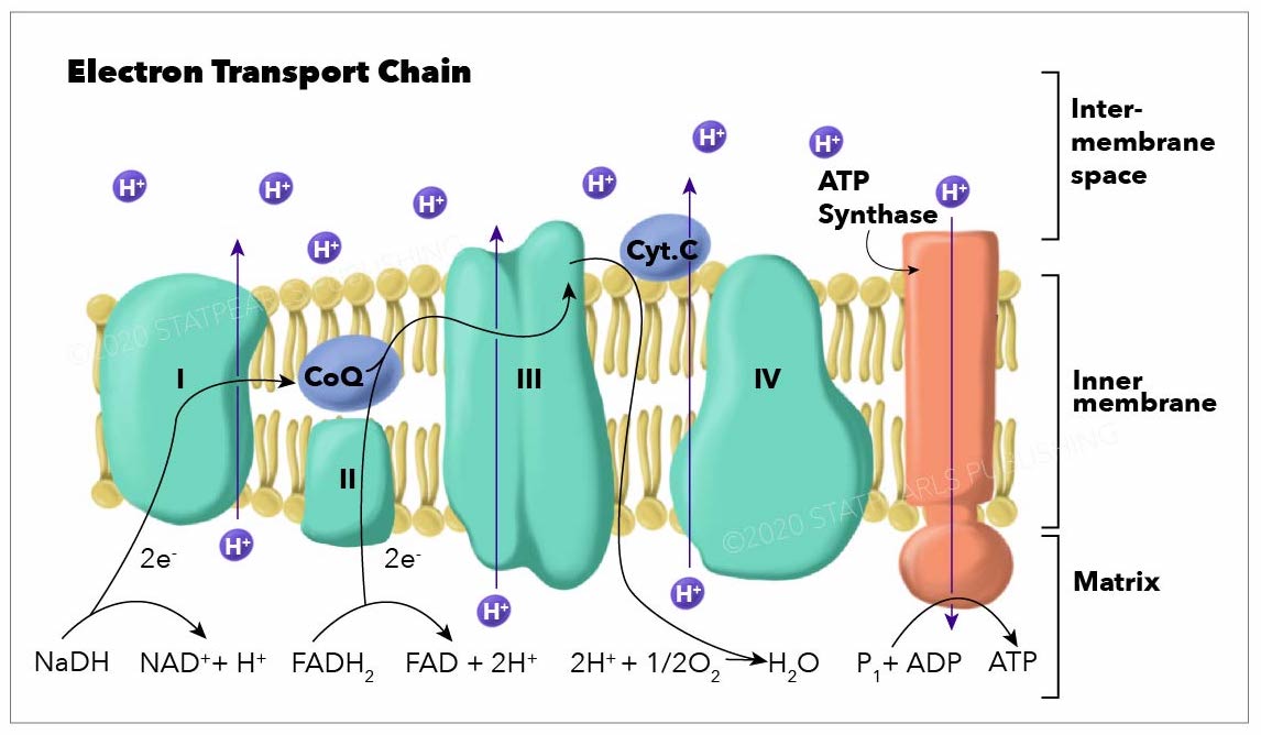
Electron Transport Concatenation graphic. Shows Inter-membrane space, inner membrane and matrix areas
Illustration by Emma Gregory
References
[1]
Lencina AM,Franza T,Sullivan MJ,Ulett GC,Ipe DS,Gaudu P,Gennis RB,Schurig-Briccio LA, Blazon 2 NADH Dehydrogenase Is the Only Point of Entry for Electrons into the Streptococcus agalactiae Respiratory Chain and Is a Potential Drug Target. mBio. 2018 Jul 3 [PubMed PMID: 29970468]
[ii]
Hirst J, Towards the molecular mechanism of respiratory circuitous I. The Biochemical journal. 2009 Dec 23; [PubMed PMID: 20025615]
[three]
Sazanov LA,Hinchliffe P, Structure of the hydrophilic domain of respiratory circuitous I from Thermus thermophilus. Science (New York, Due north.Y.). 2006 Mar 10; [PubMed PMID: 16469879]
[4]
Hirst J, Energy transduction by respiratory complex I--an evaluation of electric current cognition. Biochemical Society transactions. 2005 Jun; [PubMed PMID: 15916556]
[5]
Yankovskaya V,Horsefield R,Törnroth S,Luna-Chavez C,Miyoshi H,Léger C,Byrne B,Cecchini G,Iwata South, Architecture of succinate dehydrogenase and reactive oxygen species generation. Science (New York, N.Y.). 2003 Jan 31; [PubMed PMID: 12560550]
[seven]
Geertman JM,van Maris AJ,van Dijken JP,Pronk JT, Physiological and genetic technology of cytosolic redox metabolism in Saccharomyces cerevisiae for improved glycerol production. Metabolic engineering. 2006 Nov; [PubMed PMID: 16891140]
[8]
Thorpe C,Kim JJ, Structure and mechanism of activeness of the acyl-CoA dehydrogenases. FASEB journal : official publication of the Federation of American Societies for Experimental Biological science. 1995 Jun; [PubMed PMID: 7601336]
[9]
Sun C,Benlekbir S,Venkatakrishnan P,Wang Y,Hong S,Hosler J,Tajkhorshid E,Rubinstein JL,Gennis RB, Construction of the culling complex III in a supercomplex with cytochrome oxidase. Nature. 2018 May [PubMed PMID: 29695868]
[ten]
Iwata S,Lee JW,Okada One thousand,Lee JK,Iwata M,Rasmussen B,Link TA,Ramaswamy Due south,Jap BK, Complete construction of the 11-subunit bovine mitochondrial cytochrome bc1 complex. Science (New York, Northward.Y.). 1998 Jul three; [PubMed PMID: 9651245]
[xi]
Trumpower BL, The protonmotive Q cycle. Energy transduction past coupling of proton translocation to electron transfer by the cytochrome bc1 circuitous. The Journal of biological chemistry. 1990 Jul fifteen; [PubMed PMID: 2164001]
[12]
Hunte C,Palsdottir H,Trumpower BL, Protonmotive pathways and mechanisms in the cytochrome bc1 complex. FEBS letters. 2003 Jun 12; [PubMed PMID: 12788490]
[13]
Calhoun MW,Thomas JW,Gennis RB, The cytochrome oxidase superfamily of redox-driven proton pumps. Trends in biochemical sciences. 1994 Aug; [PubMed PMID: 7940677]
[xiv]
Schmidt-Rohr K, Oxygen Is the High-Energy Molecule Powering Complex Multicellular Life: Fundamental Corrections to Traditional Bioenergetics. ACS omega. 2020 Feb xi; [PubMed PMID: 32064383]
[fifteen]
Lovero D,Giordano L,Marsano RM,Sanchez-Martinez A,Boukhatmi H,Drechsler M,Oliva 1000,Whitworth AJ,Porcelli D,Caggese C, Characterization of Drosophila ATPsynC mutants as a new model of mitochondrial ATP synthase disorders. PloS ane. 2018 [PubMed PMID: 30096161]
[16]
Okuno D,Iino R,Noji H, Rotation and structure of FoF1-ATP synthase. Periodical of biochemistry. 2011 Jun; [PubMed PMID: 21524994]
[18]
Hinkle PC, P/O ratios of mitochondrial oxidative phosphorylation. Biochimica et biophysica acta. 2005 Jan 7; [PubMed PMID: 15620362]
[19]
Barrett MA,Zheng S,Roshankar G,Alsop RJ,Belanger RK,Huynh C,Kučerka Northward,Rheinstädter MC, Interaction of aspirin (acetylsalicylic acid) with lipid membranes. PloS i. 2012; [PubMed PMID: 22529913]
[twenty]
Warrick BJ,King A,Smolinske S,Thomas R,Aaron C, A 29-yr analysis of acute peak salicylate concentrations in fatalities reported to United states of america poison centers. Clinical toxicology (Philadelphia, Pa.). 2018 Sep [PubMed PMID: 29431532]
[21]
Cinti South, The adipose organ. Prostaglandins, leukotrienes, and essential fatty acids. 2005 Jul; [PubMed PMID: 15936182]
[22]
Enerbäck Southward, The origins of dark-brown adipose tissue. The New England journal of medicine. 2009 May vii; [PubMed PMID: 19420373]
[23]
Zhou W,Faraldo-Gómez JD, Membrane plasticity facilitates recognition of the inhibitor oligomycin by the mitochondrial ATP synthase rotor. Biochimica et biophysica acta. 2018 Sep [PubMed PMID: 29630891]
[24]
Kamalian L,Douglas O,Jolly C,Snoeys J,Simic D,Monshouwer G,Williams DP,Kevin Park B,Chadwick AE, The utility of HepaRG cells for bioenergetic investigation and detection of drug-induced mitochondrial toxicity. Toxicology in vitro : an international journal published in clan with BIBRA. 2018 Aug vii [PubMed PMID: 30096366]
[25]
Woods DM,Alsahaf H,Streete P,Dargan PI,Jones AL, Fatality after deliberate ingestion of the pesticide rotenone: a example study. Critical care (London, England). 2005 Jun; [PubMed PMID: 15987402]
[26]
Lupescu A,Jilani G,Zbidah M,Lang F, Induction of apoptotic erythrocyte decease by rotenone. Toxicology. 2012 Oct 28; [PubMed PMID: 22727881]
[28]
Weaver LK, Clinical practice. Carbon monoxide poisoning. The New England journal of medicine. 2009 Mar 19; [PubMed PMID: 19297574]
[29]
Sato Thou,Tamaki Yard,Hattori H,Moore CM,Tsutsumi H,Okajima H,Katsumata Y, Determination of total hemoglobin in forensic blood samples with special reference to carboxyhemoglobin assay. Forensic scientific discipline international. 1990 Nov; [PubMed PMID: 2279722]
[xxx]
Barker SJ,Tremper KK, The result of carbon monoxide inhalation on pulse oximetry and transcutaneous PO2. Anesthesiology. 1987 May; [PubMed PMID: 3578881]
[31]
Raub JA,Mathieu-Nolf M,Hampson NB,Thom SR, Carbon monoxide poisoning--a public health perspective. Toxicology. 2000 April seven; [PubMed PMID: 10771127]
[32]
Jensen P,Wilson MT,Aasa R,Malmström BG, Cyanide inhibition of cytochrome c oxidase. A rapid-freeze e.p.r. investigation. The Biochemical journal. 1984 Dec 15; [PubMed PMID: 6098268]
[33]
Shchepina LA,Pletjushkina OY,Avetisyan AV,Bakeeva LE,Fetisova EK,Izyumov DS,Saprunova VB,Vyssokikh MY,Chernyak BV,Skulachev VP, Oligomycin, inhibitor of the F0 function of H -ATP-synthase, suppresses the TNF-induced apoptosis. Oncogene. 2002 Nov 21; [PubMed PMID: 12444550]
Source: https://www.statpearls.com/ArticleLibrary/viewarticle/20982
0 Response to "what protein complex produces the proton gradient used to power the product of atp"
Postar um comentário